
Rediscover your healthy ambitions
No more guessing if your daily health regimen is working for you. You want results that get noticed.
Shop favorites
Formulating premium wellness products since 1997
-
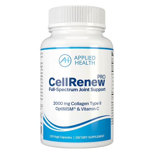
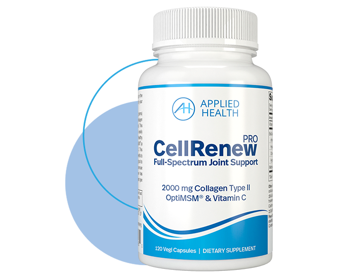
CellRenew PRO Ultimate Joint Support
Regular price $ 59.00Regular priceUnit price per -
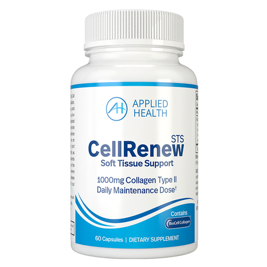
CellRenew STS Hydrolyzed Collagen Type II
Regular price $ 43.00Regular priceUnit price per -
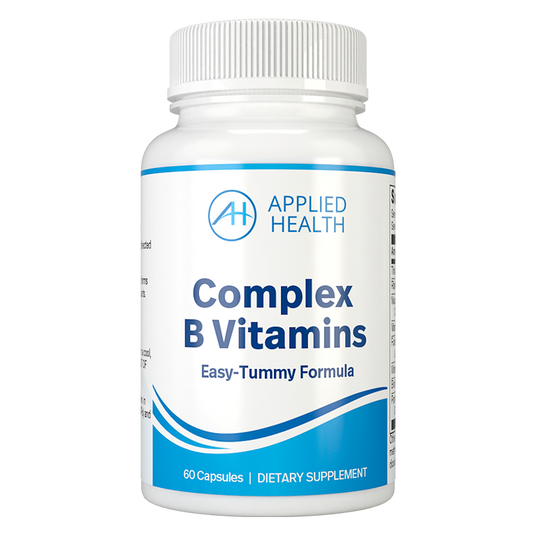
Complex B Vitamins
Regular price $ 37.00Regular priceUnit price per -
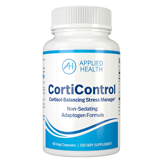
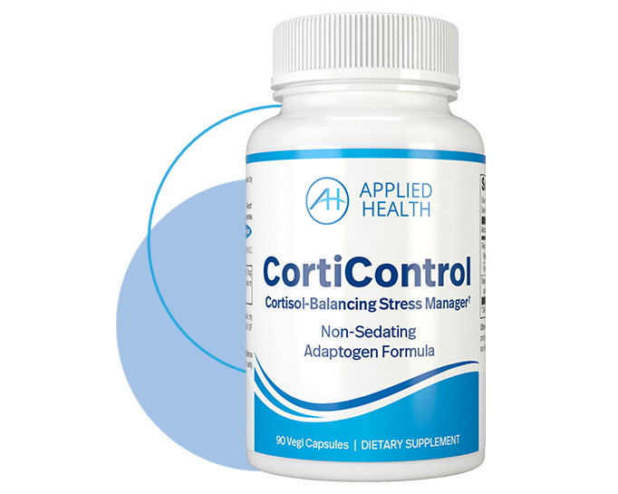
CortiControl
Regular price $ 54.00Regular priceUnit price per

Support for your Active Lifestyle
Live your life in smooth motion with health formulas designed to renew joint flexibility, energy, and active recovery.*
For all who are tough on their joints
Aches and pains keep you sitting on the sidelines? Not anymore. You have an active solution. CellRenew PRO contains 2000 milligrams of patented Collagen Type II, OptiMSM®, and Vitamin C to offer a full-spectrum joint support formula.

Join VIP Rewards
Sign up for access to product launches and insider promotions. Plus, get 10% off your first order. We will keep you in the loop — and won’t bombard your inbox.
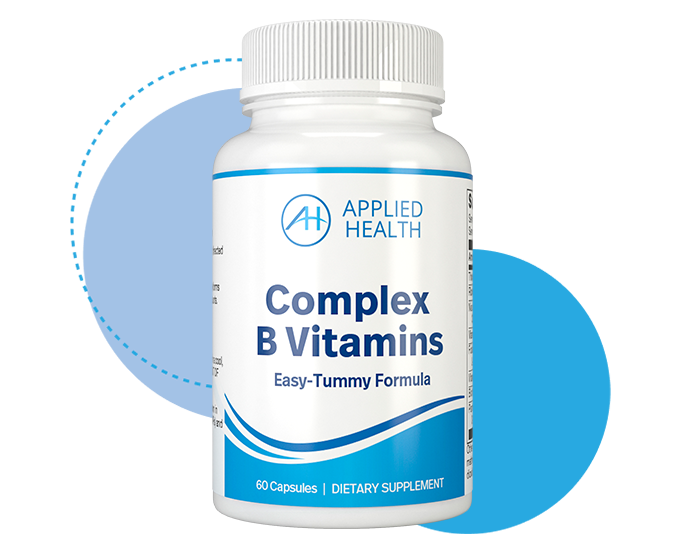
Upgrade your essential B Vitamins
Easy on your tummy. No bad afteraste. Activated B vitamin complex for optimal absorption.
Real-life experiences
See how people are achieving healthy ambitions with Applied Health.
-

Love Complex B Vitamins!
"I noticed an immediate boost in energy and stamina. I go through the day with less fatigue."*
— Tim M. —
-
My muscle aches quickly disappeared
"Soothe Ultra kept me sane as it helped lessen intense aches and soreness when I needed it most!"*
— Natalie W. —
-
Quality of life improved 1000%
"Amazing difference on my knees! I feel great with CellRenew PRO. I never want to be without it."*
— Arlene B. —

The Applied Health promise
Family-owned and operated, Applied Health was formed over 25 years ago to bring you the next-level nutrition and results that get noticed.
We only use:
● research-backed
● high-quality ingredients
● that promise predictable results.
-

None of our products contain genetically modified ingredients.
-

Our supplements are manufactured in a GMP-Certified Facility.
-

All of our products are made in the USA.


Get the healthy scoop
-
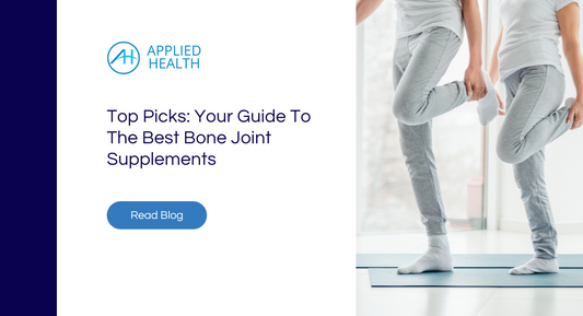
Top Picks: Your Guide To The Best Bone Joint Su...
Discover the best bone joint supplements, including glucosamine, collagen, MSM, and hyaluronic acid to enhance joint mobility and health.
Top Picks: Your Guide To The Best Bone Joint Su...
Discover the best bone joint supplements, including glucosamine, collagen, MSM, and hyaluronic acid to enhance joint mobility and health.
-
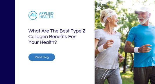
What Are The Best Type 2 Collagen Benefits For ...
Explore the amazing type 2 collagen benefits for joint, skin, and gut health. Find out which sources are best for supplementing with type 2 collagen.
What Are The Best Type 2 Collagen Benefits For ...
Explore the amazing type 2 collagen benefits for joint, skin, and gut health. Find out which sources are best for supplementing with type 2 collagen.
-

When to Start Taking Collagen Supplements
You may know the benefits of collagen, but are wondering when to start taking collagen supplements. Natural collagen production reaches its peak in your mid-20s but several factors can influence...
When to Start Taking Collagen Supplements
You may know the benefits of collagen, but are wondering when to start taking collagen supplements. Natural collagen production reaches its peak in your mid-20s but several factors can influence...
-
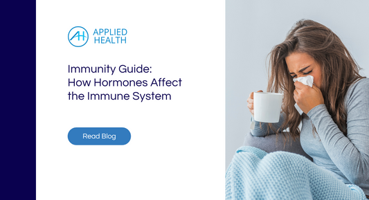
Immunity Guide: How Hormones Affect the Immune ...
Under the weather? Discover how hormones affect the immune system and which hormones have the biggest role in immunity.
Immunity Guide: How Hormones Affect the Immune ...
Under the weather? Discover how hormones affect the immune system and which hormones have the biggest role in immunity.
-

Reclaim your peace of mind — 4 stress-busting a...
Life can be a rollercoaster of emotions, and sometimes stress can take over like a clingy ex. But fear not! There are simple and effective actions you can take right...
Reclaim your peace of mind — 4 stress-busting a...
Life can be a rollercoaster of emotions, and sometimes stress can take over like a clingy ex. But fear not! There are simple and effective actions you can take right...
-
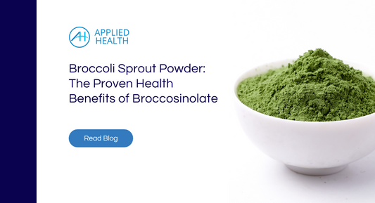
Broccoli Sprout Powder: The Proven Health Benef...
Unlock the powerful health benefits of broccoli sprout powder, rich in broccosinolate and its main phytochemical compound — sulforaphane.
Broccoli Sprout Powder: The Proven Health Benef...
Unlock the powerful health benefits of broccoli sprout powder, rich in broccosinolate and its main phytochemical compound — sulforaphane.
-
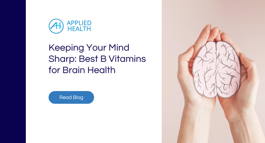
Keeping Your Mind Sharp: Best B Vitamins for Br...
Uncover the best B vitamins for brain health. Explore their roles in cognitive function and ways to prevent deficiency for optimal mental wellness.
Keeping Your Mind Sharp: Best B Vitamins for Br...
Uncover the best B vitamins for brain health. Explore their roles in cognitive function and ways to prevent deficiency for optimal mental wellness.
-
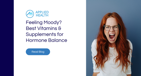
Feeling Moody? Best Vitamins and Supplements fo...
Hormone imbalances can really knock you off your feet, right? Yet, searching for the best vitamins and supplements for hormone balance can feel like a maze of information.
Feeling Moody? Best Vitamins and Supplements fo...
Hormone imbalances can really knock you off your feet, right? Yet, searching for the best vitamins and supplements for hormone balance can feel like a maze of information.


